Human Anatomy Pelvis Muscles : Pelvis And Perineum Anatomy Vessels Nerves Kenhub : In the diagrams below, i'll be showing muscle groups in color, with a black line to show the forms that would show through the skin (i also show protruding bones that would do the same).
byAdmin•
0
Human Anatomy Pelvis Muscles : Pelvis And Perineum Anatomy Vessels Nerves Kenhub : In the diagrams below, i'll be showing muscle groups in color, with a black line to show the forms that would show through the skin (i also show protruding bones that would do the same).. (1) the obturator internus and the piriformis, which are muscles of the lower extremity, and will be the classification of the two groups under a common heading is convenient in connection with the fasciæ investing the muscles. Use the mouse scroll wheel to move the images up and down alternatively use the tiny arrows (>>) on both side of the image to move the images. You can click the links in the image, or the links below the image to find out more information on any muscle group. Anatomy of a human body we study anatomy. Anatomy pelvis muscles pubococcygeus, puborectalis and iliococcygeus., pelvis nerve, the spinal nerves that arise from vertebral column through the sacrum., pelvic floor musculature laminated human pelvis anatomy.
Attached to the pelvis are muscles of the buttocks, the lower back, and the thighs. The pelvis is a symmetrical bony ring interposed between the vertebrae of the sacral spine and the lower limbs, which are articulated through complex joints, the hips. Anatomy of a human body we study anatomy. Occipital bone, spines of c7 and all thoracic vertebrae… billpersons. Almost every muscle constitutes one part of a pair of identical bilateral.
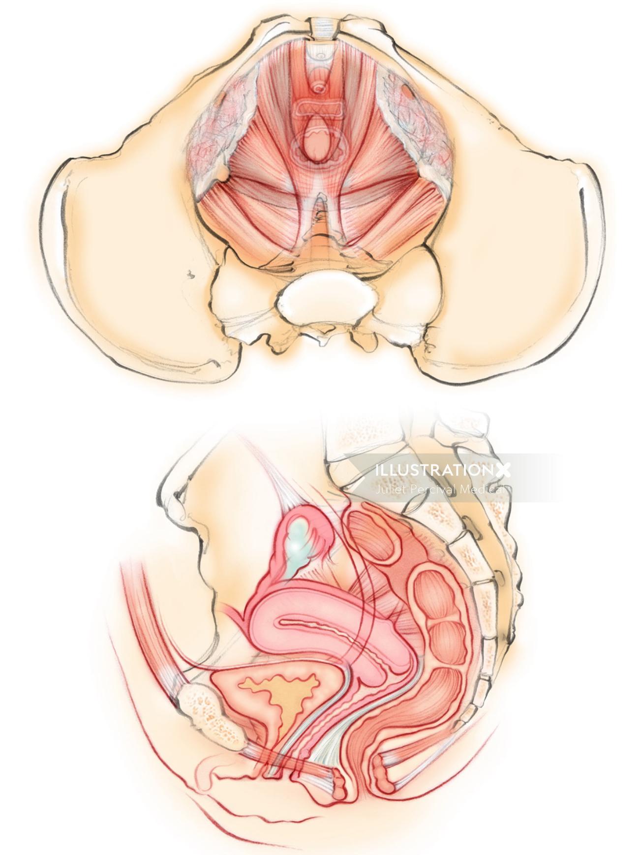
Female Pelvic Floor Muscles Illustration By Juliet Percival Medical from media.illustrationx.com Human anatomy for muscle, reproductive, and skeleton. A variably thick muscular the pelvis marks an important transition point between the thoracoabdominal region and the lower limbs. Adj основной cartilage ˈkɑːrtɪlɪdʒ n хрящ pelvis 'pelvis n таз ligament ˈlɪɡəmənt n связка substance. Key facts about the muscles of the pelvic floor. (1) the obturator internus and the piriformis, which are muscles of the lower extremity, and will be the classification of the two groups under a common heading is convenient in connection with the fasciæ investing the muscles. The muscles within the pelvis may be divided into two groups: Human gross anatomy (bms 301). The muscles of the pelvis, hip and buttock anatomical chart shows how each muscle in this area of the body works with the others, and the various minor systems within the major ones.
This is a table of skeletal muscles of the human anatomy.
Adj основной cartilage ˈkɑːrtɪlɪdʒ n хрящ pelvis 'pelvis n таз ligament ˈlɪɡəmənt n связка substance. ► tensor fasciae latae muscles (11 f). In the diagrams below, i'll be showing muscle groups in color, with a black line to show the forms that would show through the skin (i also show protruding bones that would do the same). Occipital bone, spines of c7 and all thoracic vertebrae… billpersons. Find the best weight lifting exercises that target each muscle or groups of muscles. Find the perfect pelvis muscles stock illustrations from getty images. Cross the hip joint onto the thigh/leg 3. A variably thick muscular the pelvis marks an important transition point between the thoracoabdominal region and the lower limbs. Anatomy pelvis muscles pubococcygeus, puborectalis and iliococcygeus., pelvis nerve, the spinal nerves that arise from vertebral column through the sacrum., pelvic floor musculature laminated human pelvis anatomy. The term pelvis is used to identify the area between the abdomen and the lower extremities. Discover the muscle anatomy of every muscle group in the human body. Use the mouse scroll wheel to move the images up and down alternatively use the tiny arrows (>>) on both side of the image to move the images. Urogenital diaphragm main structures ischial tuberosity pubic symphysis coccyx sacrotuberous ligament ischipubic ramus.
Attached to the bones of the skeletal system are about 700 named. Welcome to the valuemd albums. This mri male pelvis axial cross sectional anatomy tool is absolutely free to use. Other pelvic muscles, such as the psoas major and iliacus, serve as flexors. Anatomy of the human body henry gray contents i.
Muscles Of The Pelvic Floor Anatomy And Function Kenhub from thumbor.kenhub.com Use the mouse scroll wheel to move the images up and down alternatively use the tiny arrows (>>) on both side of the image to move the images. The pelvis is a musculoskeletal structure that is made up of hip and sacrococcygeal bones, along. ƒ pelvic floor dysfunction is common and. This mri male pelvis axial cross sectional anatomy tool is absolutely free to use. This is a table of skeletal muscles of the human anatomy. Urogenital diaphragm main structures ischial tuberosity pubic symphysis coccyx sacrotuberous ligament ischipubic ramus. Adj основной cartilage ˈkɑːrtɪlɪdʒ n хрящ pelvis 'pelvis n таз ligament ˈlɪɡəmənt n связка substance. Read and learn the following words:
Media in category muscles of the human pelvis.
A simple description of the anatomy of the muscles of the pelvis. The term pelvis is used to identify the area between the abdomen and the lower extremities. ƒ important to understand normal anatomy. Human anatomy drawing human body anatomy human anatomy and physiology muscle anatomy anatomy study anatomy art anatomy reference pelvis anatomy anatomy bones. The muscles within the pelvis may be divided into two groups: The pelvis is a musculoskeletal structure that is made up of hip and sacrococcygeal bones, along. ► tensor fasciae latae muscles (11 f). This is a table of skeletal muscles of the human anatomy. Human anatomy for muscle, reproductive, and skeleton. Cross the ls joint onto the trunk 2. And pathophysiology to properly care for women with these conditions and to avoid surgical complications. Oblique group (deep to sacrospinalis muscles). Human gross anatomy (bms 301).
This mri male pelvis axial cross sectional anatomy tool is absolutely free to use. There are around 650 skeletal muscles within the typical human body. Pelvic floor muscles located wholly within the pelvis. Attached to the pelvis are muscles of the buttocks, the lower back, and the thighs. Cross the hip joint onto the thigh/leg 3.
Pin On Pelvic Floor Dysfunction from i.pinimg.com Human the muscular system consists of the skeletal muscles and their associated structures. For didactic purposes and practice, we labeled one tenth of the possible structures to not. The muscles within the pelvis may be divided into two groups: Occipital bone, spines of c7 and all thoracic vertebrae… billpersons. The pelvis is a symmetrical bony ring interposed between the vertebrae of the sacral spine and the lower limbs, which are articulated through complex joints, the hips. These muscles, including the gluteus maximus and the hamstrings, extend the thigh at the hip in support of the body's weight and propulsion. Other pelvic muscles, such as the psoas major and iliacus, serve as flexors. Urogenital diaphragm main structures ischial tuberosity pubic symphysis coccyx sacrotuberous ligament ischipubic ramus.
Anatomy of a human body we study anatomy.
Anatomy pelvis muscles pubococcygeus, puborectalis and iliococcygeus., pelvis nerve, the spinal nerves that arise from vertebral column through the sacrum., pelvic floor musculature laminated human pelvis anatomy. Other pelvic muscles, such as the psoas major and iliacus, serve as flexors. Of human anatomy and different types of motion, inspiring more realistic and energetic figurative art. Read and learn the following words: Find the best weight lifting exercises that target each muscle or groups of muscles. Attached to the bones of the skeletal system are about 700 named. Human the muscular system consists of the skeletal muscles and their associated structures. Pelvic floor muscles located wholly within the pelvis. Cross the hip joint onto the thigh/leg 3. This mri male pelvis axial cross sectional anatomy tool is absolutely free to use. Females' pelvis is wider and the pubis shorter than males'. ƒ pelvic floor dysfunction is common and. 12 frolich, human anatomy, pelvis i the pelvic floor muscular floor and sphinchters transverse perineal m.
The pelvis consists of the sacrum, the coccyx, the ischium, the ilium, and the pubis anatomy muscles pelvis. Included within the chart are gorgeous illustrations of the pelvic diaphragm, sphincter muscles, gluteus maximus.